X-ray Diagnosis – AI-Powered Image-to-Text Analysis
Hi! Today, I’m excited to present our Proof of Concept (PoC) for diagnosing chest X-rays.
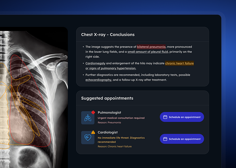
The system detects and localizes lesions on chest X-rays. It’s designed to provide patients with a detailed diagnosis presented in easy-to-read text, along with interactive visualizations that highlight the areas where lesions are detected. By outlining regions of interest and summarizing key findings, the system enhances readability and helps patients better understand their results.
To begin, the user uploads an X-ray file. The system supports various formats, including DICOM, PDF, PNG, and JPEG.
The X-ray scan is processed, and the system provides a diagnosis based on the image.
The diagnosis is divided into three sections: Conclusions, Suggested Appointments, and Detailed Diagnosis.
The user can hover over the section dedicated to lung fields, where the system highlights lesions on the X-ray that correspond to descriptions in the text.
Each color indicates the severity of the lesion and the immediacy of the threat.
___________________
We are Autentika
UX-Driven Design & Development Agency that for more than 15 years has been building top-quality web, e-commerce, and mobile apps.
They call us "the agency for demanding projects." The truth is that we like challenges and get satisfaction from well-done work.
Check us out at www.autentika.com
Follow us:


